Diagnostic Imaging Referrals in Stouffville: X-Ray, Ultrasound, CT, and MRI Explained
Need a scan? How Ontario imaging requisitions work, how X-ray, ultrasound, CT and MRI differ, how to prepare, and how Stouffville patients get their results.
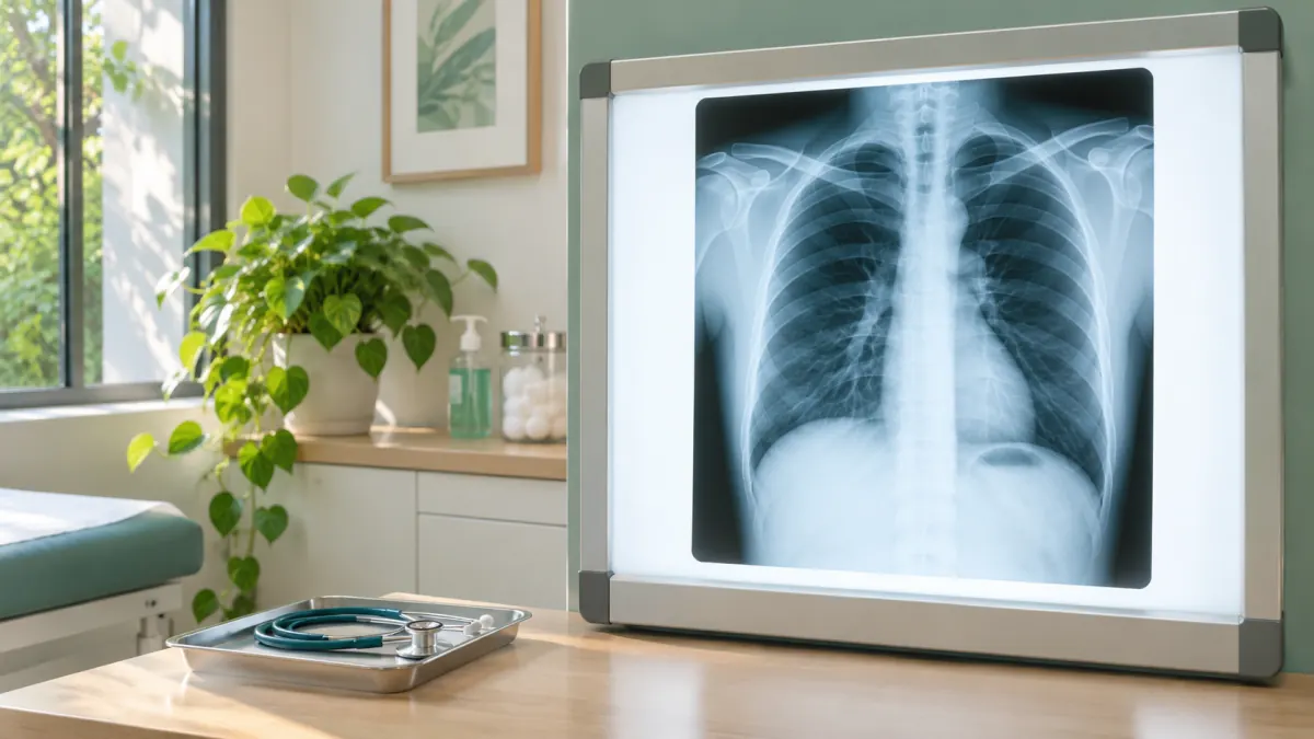
An X-ray of a sprained wrist can be over before you have finished taking off your coat. An MRI of the same wrist might mean lying very still inside a humming tunnel for half an hour or more. Both are “imaging,” both start with a piece of paper from your clinician, and both end with a report that goes back to the person who ordered it. Beyond that, they could hardly be more different, and knowing which one you are heading for, and why, makes the whole process less mysterious.
In Ontario, medical imaging such as X-ray, ultrasound, CT, and MRI requires a requisition from a clinician. You cannot simply book one yourself, with the notable exception of screening mammograms for eligible people through the Ontario Breast Screening Program. This guide walks Stouffville patients through how imaging referrals work, what each type of test is good at, how to prepare, and what happens after the pictures are taken.
From symptom to scan: how the process works
- An appointment comes first. A clinician assesses your symptoms, examines you, and decides whether imaging would actually help answer a question. Sometimes the answer is no, and that is a good thing; more on that below.
- The requisition is completed. It lists the test, the body part, the clinical question, and how urgent it is. The clinical information helps the radiologist choose the right technique and interpret the images.
- The appointment is booked. Depending on the test and local arrangements, either the clinic sends the requisition to an imaging provider, or you receive the requisition and call to book. Some tests are done at community imaging clinics, while others are typically done in hospitals. Your clinic will tell you which applies.
- You prepare, if needed. Some tests need no preparation at all; others need fasting, a full bladder, or a review of kidney function or implants.
- The images are taken by a trained technologist or sonographer.
- A radiologist interprets the images, which are reviewed by a doctor who specializes in reading them, and a written report is sent to the clinician who ordered the test.
- Your clinician follows up to explain the results and next steps.
If you are unsure where you are in that process, call the office that ordered the test rather than the imaging provider. Our diagnostic imaging referrals page has a short summary as well.
The main types of imaging, compared
| Test | How it works | Commonly used for | Radiation? | Typical length |
|---|---|---|---|---|
| X-ray | A small dose of radiation passes through the body to create a picture | Bones, fractures, chest and lungs, some joint problems | Yes, usually a low dose | Minutes |
| Ultrasound | High-frequency sound waves bounce off tissues | Abdomen, pelvis, thyroid, breast lumps, blood vessels, pregnancy, soft tissue lumps | No | 15 to 45 minutes |
| CT (computed tomography) | Many X-ray images combined by computer into detailed cross-sections | Detailed views of the chest, abdomen, head, kidney stones, complex fractures | Yes, more than a plain X-ray | Usually under 30 minutes, including setup |
| MRI (magnetic resonance imaging) | Strong magnets and radio waves create detailed images | Brain, spine, joints, ligaments, soft tissues | No | Often 30 to 60 minutes |
Other imaging tests you may hear about include mammography for breast imaging, bone density scans to check for osteoporosis, and nuclear medicine scans, which use small amounts of radioactive tracers to show how organs are working.
X-ray
The workhorse of imaging. X-rays are fast, widely available, and excellent for bones. A chest X-ray can show pneumonia, fluid, or an enlarged heart. Preparation is minimal: you may be asked to change into a gown and remove jewellery or anything metal in the area being imaged.
Ultrasound
Ultrasound uses no radiation, which makes it the first choice for pregnancy and a common first step for many abdominal, pelvic, and neck concerns. A gel is applied to the skin, and a handheld probe is moved over the area. Some ultrasounds are done internally, such as a transvaginal pelvic ultrasound, which you will be told about beforehand. Preparation varies:
- Abdominal ultrasound often requires fasting for several hours beforehand, so the gallbladder and other organs can be seen clearly.
- Pelvic ultrasound often requires a full bladder, which means drinking a specified amount of water ahead of time and not emptying your bladder until after the scan.
CT
CT provides detailed cross-sectional images and is invaluable in many situations. Some CT scans use an iodine-based contrast dye, given by IV or by mouth, to highlight blood vessels and organs. If contrast is planned, you may be asked about kidney function, diabetes medications, and previous reactions to contrast. Because CT uses more radiation than a plain X-ray, it is ordered when the benefit clearly outweighs the small added risk.
MRI
MRI produces remarkably detailed images of soft tissues like the brain, spinal cord, cartilage, and ligaments, without radiation. The strong magnet means careful safety screening:
- You will be asked about pacemakers, defibrillators, cochlear implants, metal clips, joint replacements, shrapnel, or work involving metal fragments.
- Some implants are safe for MRI; others are not or require special settings. Bring any implant cards you have.
- The machine is noisy, so earplugs or headphones are provided.
- If you are claustrophobic, mention it to your clinician ahead of time. Some people find strategies or a mild medication helpful, and you would then need someone to drive you home.
Some MRI scans also use a contrast agent given by IV.
Why your clinician may not order a scan
It is natural to want a picture of what is going on. But imaging is not always the best next step, and more scans do not always mean better care. A few examples:
- Low back pain without red flags usually improves within weeks, and early imaging often shows age-related changes that are normal and unrelated to the pain. Imaging is generally reserved for back pain with warning signs or pain that is not improving as expected.
- Most headaches do not need a brain scan unless there are specific concerning features.
- Minor ankle and foot injuries can often be assessed with clinical rules that show whether an X-ray is likely to be useful.
Unnecessary imaging can lead to incidental findings, harmless spots or variations that trigger more tests and worry, and some imaging adds radiation exposure. If your clinician suggests waiting, it is fair to ask what would change that plan, and what to watch for in the meantime.
How long will I wait?
Wait times vary by test type, urgency, and the provider. X-rays are often available quickly, while non-urgent CT and MRI scans can take longer. Your clinician indicates urgency on the requisition, and radiology teams prioritize based on medical need. If your symptoms get worse while you are waiting, contact your clinician; they may be able to update the request or arrange a different plan.
For true emergencies, such as a suspected stroke, serious head injury, sudden severe abdominal pain, or chest pain, do not wait for an outpatient scan. Call 911 or go to the nearest emergency department. For Stouffville residents, that is usually Markham Stouffville Hospital or Uxbridge Hospital.
What to bring and how to prepare
- Your Ontario health card
- Your requisition, if you were given a paper copy
- A list of your medications and allergies, especially any previous reaction to contrast dye
- Implant cards or information about any metal in your body, for MRI
- Details of previous imaging done elsewhere, so the radiologist can compare
- Comfortable clothing without metal zippers or buttons
- Follow the specific preparation instructions you are given about fasting, bladder filling, or medications
Pregnant or possibly pregnant? Tell your clinician and the imaging staff before any X-ray, CT, or nuclear medicine test. Ultrasound and, in many cases, MRI can be used instead.
Getting your results
Results are sent to the clinician who ordered the test, not usually directly to the patient first. Some imaging providers also offer access through online patient portals, but the report is written in specialized language, and a phrase that looks alarming may be completely routine. Your clinician will put it in context.
A few tips:
- Ask when and how you will hear about results at the time the test is ordered. Some clinics call only if follow-up is needed, while others book a follow-up appointment to review every result.
- Do not assume no news is good news indefinitely. If you have not heard anything within the expected time, call the clinic.
- Expect possible next steps. Results sometimes lead to further imaging, blood work, or a specialist referral. If lab tests are part of the plan, our post on blood work in Stouffville explains how that side of things works.
OHIP and imaging
Medically necessary imaging ordered by a clinician is generally covered by OHIP for eligible Ontario residents with a valid health card. Imaging requested for non-medical reasons, such as some third-party requirements, may not be covered by OHIP. If cost is a question for you, ask before your test.
Questions about imaging
Can I ask my doctor for an MRI instead of an X-ray?
Is the radiation from an X-ray or CT dangerous?
My requisition is from a walk-in visit. Who follows up?
Arranging imaging through HealthSmart
If you have a symptom that might need imaging, start with an assessment. You can book an appointment at our clinic in Stouffville, and if a test is appropriate, we will prepare the requisition and explain the next steps. If you are waiting on results from a test we ordered, please get in touch and our team will help. Many Stouffville families find the process simpler once they know what to expect.
This article provides general information about imaging and is not a substitute for individual advice from the clinician managing your care.